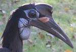
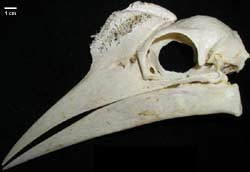
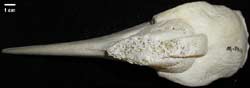
The hornbills comprise Bucerotiformes, which is divided into two distinct groups, Bucerotidae and the Bucorvidae (ground hornbills), all of which are easily distinguished from other birds by the large casque that sits on top of their skull. |
|
|
This distinctive feature is formed by the expansion of the posterior end of the upper mandible and is comprised of a bony core at the base that may be either highly cancellated (as in Bucorvus) or more solid. A keratinous sheath overlies the bony base of the casque. The function of the casque is debatable, it likely is used as a identification device for hornbills, imparting such information as species, sex, and age. The hollow nature of the casque may also be utilized to amplify communications between hornbills (Alexander et al., 1994). |
|
The closest relatives of the hornbills reportedly are the whoopoes, woodhoopoes, and scimitarbills (Upupiformes; Sibley and Alquist, 1991). Hornbills are found only in the Old World from Africa to India, the Malaysian archipelago, New Guinea, and the Solomon Islands. Despite their expansive distribution, their fossil record is sparse. Up to this time, hornbill fossils date back as far as the Mid-Miocene of Morrocco, but the only material that has been identified with any certainty is attributed to the ground hornbills (Olson, 1985).
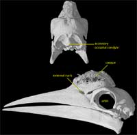
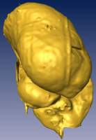
Bucerotiforms are easily distinguished from other birds not only by their large casque, but also by a number of other distinctive features. Other skeletal characters include the fusion of the atlantal and axial vertebrae and the possession of an accessory supraoccipital condyle in addition to the basioccipital condyle (this feature is seen easily in the 3D reconstructions of the skull of Bucorvus abyssinicus). The buceriforms also lack a middle lobe to their kidney (Johnson, 1979) and possess long, flattened eyelashes (Kemp, 1995). |
|
|
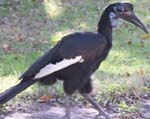
Bucorvus abyssinicus, the northern ground hornbill, and B. leadbeateri, the southern ground hornbill, are the sole members of Bucorvidae, the most basally diverging taxon within Bucerotiformes. They are distinguished from other bucerotiforms by the possession of 15 (instead of 14) cervical vertebrae, elongated tarsi, the absence of the cranial carotid arteries, and by not sealing the entrance to their nest cavity (Kemp, 1995). |
|
Both species of ground hornbills live in Africa; however, B. abyssinicus inhabits the northern portion of sub-Saharan Africa south to the equator while the range of B. leadbeateri extends from the equator to the southern tip of the continent. Both species inhabit savannah and grassland-type environments, and their ranges actually overlap in Kenya. The two ground hornbills are incredibly similar in appearance, but B. abyssinicus is distinguished from B. leadbeateri by a higher casque with three lobes and a large yellow patch at its base (Kemp, 1995).
Buvorvus abyssinicus typically travels in pairs, trios, or quartets but breeds in pairs. They are primarily carnivorous birds, consuming small organisms such as tortoises, lizards, amphibians, mammals, other birds, and insects (although they may supplement their diet with nuts, fruit, and seeds). The northern ground hornbill enjoys fairly stable numbers; although the numbers of its southern relative are declining rapidly due to the development of their habitat (Kemp, 1995).

Literature
Alexander, G. D., D. C. Houston, and M. Campbell. 1994. A possible acoustic function for the casque structure in hornbills (Bucerotidae). Journal of Zoology, London, 233:57-67.
Baumel, J. J., A. S. King, J. E. Breazile, H. E. Evans, and J. C. Vanden Berge (eds.). 1993. Handbook of Avian Anatomy: Nomina Anatomica Avium, Second Edition. Publication of the Nuttall Ornithological Club, number 23. Nuttall Ornithological Club, Cambridge, Massachusetts, 779 pp.
Beddard, F. E. 1889. Contributions to the anatomy of Picarian birds. Part I. On some points in the structure of hornbills. Proceedings of teh Zoological Society of London 1889:587-594.
Beddard, F. E. 1901. Notes on the anatomy of Picarian birds. No. IV. On the skeletons of Bucorvus cafer and Bucorvus abyssinicus with notes on other hornbills. Proceedings of the Zoological Society of London 1:16-24.
Burton, P. J. K. 1984. Anatomy and evolution of feeding apparatus in the avian orders Coraciiformes and Piciformes. Bulletin of the British Museum (Natural History), Zoology 47:331-443
Elliott, D. G. 1878. Bucorvus: remarks on the genus, with figure of heads of Bucorvus abyssinicus, Bucorvus guineensis, Bucorvus cafer, and Bucorvus pyrrhopsis. Bulletin de la Societe Zoologique de France 3:34-36.
Garrod, A. H. 1876. Bucorvus abyssinicus, some peculiarities in its anatomy described. Proceedings of the Zoological Society of London 1876:60.
Johnson, O. W. 1979. Urinary organs; pp. 183-236 in A. S. King and J. McLelland (eds.), Form and Function in Birds, Volume 1. Academic Press, London.
Kemp, A. C. 1995. The Hornbills. Oxford University Press, Oxford, 302 pp.
Kemp, A. C., and Kemp, M. I. 1980. The biology of the southern ground hornbill, Bucorvus leadbeateri (Aves, Bucerotidae). Annals of the Transvaal Museum 32:65-100.
Lipscomb, C. 1948. Nesting of Bucorvus abyssinicus. Ibis 14:153-154.
Olson, S. L. 1985. The fossil record of birds; pp. 136-138 in D. S. Farner, J. R. King, and K. C. Parkes (eds.), Avian Biology, Vol. 8. Academic Press, London.
Ottley, W. 1879. Bucorvus abyssinicus, on the vessels in its head and neck. Proceedings of the Zoological Society of London 1879:461-467.
Sibley, C. G., and J. E. Ahlquist. 1991. Phylogeny and Classification of Birds: A Study in Molecular Evolution. Yale University Press, New Haven, Connecticut, 976 pp.
Links
Bucorvus abyssinicus on the Fort Worth Zoo website, WhoZoo.
Images of hornbills at the Natural Encounters Inc. website.


 )