 Expert annotations for this species! See below. Expert annotations for this species! See below.
The Pacific Giant Salamander, Dicamptodon ensatus, and its close relatives, are the largest and most massive of the salamanders that have a terrestrial stage. This species occupies a small region of California, in the Coast Range west of San Francisco Bay from southern Santa Cruz County to southern Mendocino County, where it is replaced to the north by a close relative, D. tenebrosus. | |
Giant salamanders have an interesting life history. They lay large clutches of sizable, yolky eggs under rocks in the headwaters of clear water streams. Eggs hatch after a several month period of incubation into stream-adapted larvae that have well developed limbs, a depressed body, and a tail fin that extends only to the base of the tail. The larvae live for at least two and perhaps as long as four years, or even more. They grow to large size, some as large as the largest known adults. Occasionally larvae become reproductive, but more typically they metamorphose into the adult form. The skull shown here is from a large adult.
The head of D. ensatus is massive for a terrestrial salamander, with a robustly developed skull comprised of relatively stout bones that are well articulated to each other. Key features are illustrated below.
Dorsal View
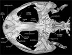 |
Dicamptodon ensatus, dorsal view. |
|
In dorsal view note the large orbital vacuities which contain the eyes. In front of them is the face and jaws -- the premaxillae, septomaxillae, nasals, lacrimals, prefrontals, frontals, parietal, and maxillae -- and behind them is the occipital and otic region. Click on the thumbnail for a detailed description of the skull in dorsal view.
|
Ventral View
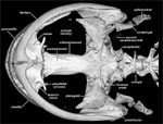 |
Dicamptodon ensatus, ventral view. |
|
In ventral view the dentary, vomers, parasphenoid, orbitosphenoid, pterygoids, and hyobranchial skeleton can be seen. The hyobranchial skeleton is an important part of the head of salamanders and provides the skeleton of the tongue. Click on the thumbnail for a detailed description of the skull in ventral view.
|
Sagittal Cutaway View
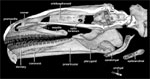 | Dicamptodon ensatus, sagittal cutaway. |
|
The lower jaw includes only two bones in most salamanders, but in Dicamptodon there are three. Click on the thumbnail to learn more about the lower jaw and to see the internal surface of other bones. |

About the Species
This specimen was collected from Empire Cave, Santa Cruz County, California by R. E. Graham in 1960. It was made available to the University of Texas High-Resolution X-ray CT Facility for scanning by Dr. David Wake of the University of California, Berkeley. Funding for scanning was provided by a National Science Foundation Digital Libraries Initiative grant to Dr. Timothy Rowe of The University of Texas at Austin.
About this Specimen
The specimen was scanned by Matthew Colbert on 21 November 2001 along the coronal axis for a total of 404 1024x1024 pixel slices. Each slice is 0.089 mm thick, with an interslice spacing of 0.089 mm and a field of reconstruction of 35 mm.
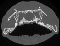 |
To the left is a reduced sample slice through the skull of Dicamptodon. Click on the thumbnail to view an unreduced version. |
About the
Scan
Literature
Rainer, R. 1998. Homology of cranial ossifications in urodeles: significance of developmental data for fossil basal tetrapods. Neues Jahrbuch für Geologie und Paläontologie Monatshefte 1998:1-25.
Links
Dicamptodon ensatus on AmphibiaWeb (University of California, Berkeley)
Dicamptodon ensatus on The Animal Diversity Web (The University of Michigan Museum of Zoology)
Dicamptodontidae on the Tree of Life (University of Arizona)
Literature
& Links
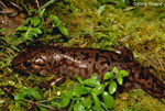
Front page image.

|
Additional Imagery
 |


 )


)
)