Throughout trilobite evolution, various clades independently converged upon morphologies that permitted enrollment of their exoskeletons so as to efficiently encase their soft tissues within a hard protective carapace (Bergström, 1973; Esteve et al., 2011). While the general advantages of this defensive posture are intuitively obvious, little is yet known about the material and biomechanical conditions of the exoskeleton that are necessary for enrollment to be an effective protective strategy. Three-dimensional models of their exoskeletons may allow both qualitative and quantitative assessments of the protective capabilities of trilobites and may reveal important constructional and functional parameters that are necessary for enrollment to be effective against predation, with broader implications for predator-prey escalation and evolution.
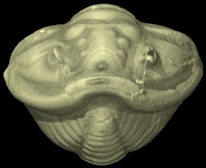
Derived trilobites, such as the Late Ordovician Flexicalymene meeki shown here, had differentially-thickened cuticles (between 200 and 400 microns) and a number of coaptative devices, morphological structures that guided the articulation between segments and locked the exoskeleton in an enrolled posture. Flexicalymene meeki exhibits beveled edges on the tips of the pleurae, dorsally-positioned fulcra, and crescent-shaped anterior extensions of the axis at each thoracic segment, all of which constrained the articulation and provided structural support between sclerites during enrollment. Some trilobites use additional coaptative devices, such as sockets along the cephalic doublure (ventral rim of the cephalon) into which the tips of the pleurae and the pygidium (tail) can be inserted to provide resistance to shearing predators. However, F. meeki employs a different strategy marked by the insertion of the posterior tip of the pygidium into a small ventral cavity near the anterior of the cephalon. This style of enrollment may have provided both shearing and compressive resistance.
Initial Finite Element analyses on the segmented digital model of the trilobite indicate that differential thickening of the cuticle evidently increased the compressive resistance of the exoskeleton. The imbricated tips of the pleurae may have served to laterally thicken the overall morphology during enrollment, providing increased resistance to shearing and laterally-directed compressive forces that may have occurred during a predatory attack.

About the Species
This specimen was collected from the late Ordovician of Leesburg, Ohio. It was made available to The University of Texas High-Resolution X-ray CT Facility for scanning by Dr. Nigel Hughes and Mr. Mathew Knauss of the University of California Riverside. Funding for scanning and image processing was provided by UTCT.
About this Specimen
The specimen was scanned by Jessie Maisano on 18 February 2014 along the coronal axis for a total of 764 slices. Voxel size is 0.020 mm.
About the
Scan
Literature
Bergström, J. 1973. Organization, life, and systematics of trilobites. Fossils and Strata 2:1-69.
Esteve, J., Hughes, N.C., Zamora, S. 2011. Purujosa trilobite assemblage and the evolution of trilobite enrollment. Geology 39:575-578.
Literature
& Links
Front page image.
|  |
Additional
Imagery
|


 ,
,  )


)