Excerpted from Evans, S.E., C. Lally, D.C. Chure, A. Elder, and J.A. Maisano. 2005. A Late Jurassic salamander (Amphibia: Caudata) from the Morrison Formation of North America. Zoological Journal of the Linnean Society 143:599-616.
Amphibia, Caudata, Urodela
Iridotriton hechti Evans et al., 2005
Specimen:
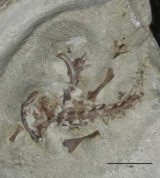
DINO 16453 is a small specimen preserving parts of a single skeleton missing only small skull bones, digits and the distal tail. This is the type and only specimen. It was recovered at the Rainbow Park microsite (Dinosaur National Monument No. 96), Utah, USA, and is held in the collections of Dinosaur National Monument. The horizon from which it came is the Brushy Basin Member of the Morrison Formation, Upper Jurassic (c.150-148 Myr, Kowallis et al., 1998: Kimmeridgian or early Tithonian in age). The name Irido triton comes from two words: iris, irido meaning rainbow (an allusion to the Rainbow Park site); and triton, meaning newt. The species name recognises Max Hecht, formerly of the American Museum of Natural History, New York, who was the first person to describe salamander material from the Morrison Formation.
General features:
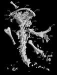
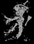
The specimen preserves a small (snout-sacrum length c.55 mm) fully metamorphosed salamander. DINO 16453 remains in articulation and includes much of the skull, the complete presacral axial skeleton, the sacrum, a small set of postsacrals, and parts of the girdles and the limbs. The skull, forelimbs, and anterior presacral series (DINO 16453a) are preserved in dorsal view, but the posterior presacral region and left hind limb are on a small block (DINO 16453b) detached during collection and prepared in ventral view. Only the anterior block was scanned and is shown in the images here.
The specimen is generally well preserved in three dimensions, with the vertebrae fully articulated, but there has been some disarticulation of the limbs and girdles and of parts from the right side of the skull roof and jaws. It is not possible to get an accurate measurement of the snout-sacral length, but comparison of humeral and femoral lengths with those of similarly proportioned modern analogues suggests a snout-sacrum length of 50-60mm, and a total length (with tail) of between 80-100mm overall. Despite this small size, the specimen appears to represent a metamorphosed individual (dermal roofing bones ossified and in position; squamosal full size; all bones of lower jaw ossified; dorsal process of maxilla ossified; vomer fully formed; otic capsule complete and stapes ossified [Rose, 2003]).
The following combination of characters distinguishes Iridotriton hechti from other fossil and modern salamanders: thin unsculptured skull bones; a fully open Meckelian fossa in the dentary; premaxilla with wide, short alary process having angled lateral edge; prootic, opisthotic and exoccipital form a single unit, although sutural lines separate the opisthotic from the other bones; stapes free; parasphenoid without internal carotid foramina, narrower anteriorly than posteriorly; an estimated 17 presacral vertebrae; simple ectochordal vertebral centra, with small anterior basapophyses but no ventromedian keel; atlas shorter than succeeding vertebrae; spinal nerve foramina in atlas and in tail vertebrae; co-ossified scapula and coracoid, with narrow, waisted scapula and large, heavily ossified coracoid plate perforated by supracoracoid foramen; strongly built forelimbs; radius with expanded distal head; well-ossified tarsus and carpus including fusion of ulnare and intermedium; rib bearers with conjoined heads throughout the column.
Crown group salamanders are currently divided into two groups, Cryptobranchoidea (including cryptobranchids and hynobiids) and Salamandroidea (all remaining salamanders). The balance of characters suggests a position for Iridotriton on the stem of salamandroid salamanders.
Specimen description:
Click on the thumbnails below for a detailed description of the specimen in dorsal and ventral views.

Literature
Cannatella DC, Hillis DM. 1993. Amphibian relationships: phylogenetic analysis of morphology and molecules. In: Cannatella DC, Hillis DM, eds. Amphibian relationships. Phylogenetic analysis of morphology and molecules. Herpetological Monographs 7: 1-7.
Dollo L. 1884. Note sur le batracien de Bernissart. Bulletin du Musée Royal dHistoire Naturelle de Belgique 3: 85-96.
Dong Z, Wang Y. 1998. A new urodele (Liaoxitriton zhongjiani gen. et sp. nov.) from the Early Cretaceous of Western Liaoning Province, China. Vertebrata PalAsiatica 36: 159-172 (In Chinese with an English summary).
Duellman WE, Trueb L. 1986. Biology of amphibians. New York, St Louis, San Francisco: McGraw-Hill. 570pp.
Edwards JL. 1976. Spinal nerves and their bearing on salamander phylogeny. Journal of Morphology 148: 305-328.
Estes R. 1981. Gymnophiona, Caudata. In: Wellnhofer P. ed. Handbuch der Paläoherpetologie 2. Stuttgart: Gustav Fischer Verlag. 115 pp.
Evans, S.E., Lally, C., Chure, D.C., Elder, A. and Maisano, J.A. 2005. A Late Jurassic
salamander (Amphibia: Caudata) from the Morrison Formation of North America.
Zoological Journal of the Linnean Society (in press).
Evans SE, Milner AR. 1996. A metamorphosed salamander from the Early Cretaceous of Las Hoyas, Spain. Philosophical Transactions of the Royal Society of London B 351: 627-646.
Evans SE, Milner AR, Mussett F. 1988. The earliest known salamanders (Amphibia, Caudata): a record from the Middle Jurassic of England. Geobios 21: 539-552.
Feller AE, Hedges SB. 1998. Molecular evidence for the early history of living amphibians. Molecular Phylogenetics and Evolution 9: 509-516.
Francis ETB. 1934. The anatomy and the salamander. Clarendon Press, Oxford, 281pp.
Gao KQ, Shubin NH. 2001. Late Jurassic salamanders from northern China. Nature 410: 574-577.
Gao K, Shubin NH. 2003. Earliest known crown-group salamanders. Nature 422: 424-428.
Hay JM, Ruvinsky I, Hedges SB, Maxson L.R. 1995. Phylogenetic relationships of amphibian families inferred from DNA sequences of mitochondrial 12S and 16S ribosomal RNA genes. Molecular Biology and Evolution 12: 928-937.
Hecht MK, Estes R. 1960. Fossil amphibians from Quarry Nine. Postilla 46: 1-19.
Kowallis BJ, Christiansen EH, Deino AL, Peterson F, Turner CE, Kunk MJ, Obradovich
JD. 1998. The age of the Morrison Formation. In: Carpenter K, Chure DJ, Kirkland JI, eds. The Upper Jurassic Morrison Formation: an interdisciplinary study. Part 1. Modern Geology (Special Issue) 22: 235-260.
Milner AR. 1983. The biogeography of salamanders in the Mesozoic and early Caenozoic: a
cladistic-vicariance model. In: Sims RW, Price JH, Whalley PES, eds. Evolution, time and space: the emergence of the biosphere. London and New York: Academic Press, 431-468.
Milner AR. 1988. The relationships and origin of living amphibians. In: Benton MJ, ed. The Phylogeny and Classification of the Tetrapods. Vol. 1. Amphibians, Reptiles, Birds. Systematics Association Special Volume, 35A. Oxford: Clarendon Press, 59-102.
Milner AR. 2000. Mesozoic and Tertiary Caudata and Albanerpetontidae. In: Heatwole H, Carroll RL eds. Amphibian Biology, Vol.4, Palaeontology.Chipping Norton, New South Wales, Australia, 1413-1444.
Rose CS. 2003. The developmental morphology of salamander skulls. In: Heathole H, Davies M, eds. Amphibian Biology, Volume 5. Osteology. Australia: Surrey Beatty and Sons Pty. Ltd., 1686-1783.
Trueb L. 1993. Patterns of cranial diversity among the Lissamphibia. In: Hanken J, Hall BK, eds. The Skull. Volume 2, Patterns of Structural and Systematic Diversity. Chicago: The University of Chicago Press, 255-343.


 ,
,  )