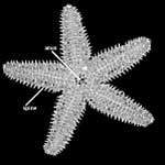
Pisaster is a genus of starfish that includes three species, P. brevispinus, P. giganteus, and P. ochraceus (Smith and Carlton, 1975). The geographic distribution of Pisaster extends along the Pacific coast from Alaska to southern California in intertidal zone habitats. Pisaster can reach up to 28 cm in size and typically has five arms, although many individuals have between four and seven arms. Individuals can vary greatly in color, most commonly including yellow, orange, brown, and purple (Hutchins et al., 2003) (the specimen shown above has been false-colored).
Sea stars belong to a group of invertebrates called Asteroidea (Lafay et al., 1995; Anderson, 2001; Brusca and Brusca, 2003). Asteroidea contains about 300 extant genera and 1600 extant species of sea stars, which are cosmopolitan in distribution (Lafay et al., 1995; Hutchins et al., 2003). Sea stars are well known for their star shape with five arms radiating from a central body. However, shape in sea stars is quite variable with some species having as many as 40 arms (Anderson, 2001).
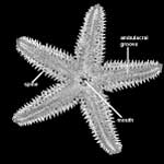
The body of sea stars has two sides; the oral side has the opening for the mouth and the aboral side (shown above) has the opening for the anus. The mouth is surrounded by connective tissue and typically a ring of protective spines. On the oral side of each arm there are two to four rows of tube feet. These feet can be retracted into a groove that runs the length of each arm (known as the ambulacral groove) when not in use and are protected by a series of spines that cover the grooves opening. In Pisaster, the feet are suckered, an adaptation for moving along hard substrates (Anderson, 2001).
The aboral surface of sea stars is covered with spines. In addition to spines, Pisaster also has structures called pedicellariae. Pedicellariea are thin structures with a jaw-like end that are used for crushing food, catching prey, and protecting the sea star from smaller organisms trying to settle on its body (Anderson 2001).
Sea stars are characterized as either opportunistic predators or scavengers. Their diet consists of many invertebrate species, including sponges, shellfish, corals, bivalves, and algae (Anderson, 2001; Brusca and Brusca, 2003; Hutchins et al., 2003). Additionally, sea stars have been observed eating dead animal matter (Brusca and Brusca, 2003). While many species digest their food internally, some species have the ability to protrude the front portion of their stomach through their mouth to ingest food that is too large to fit inside their body (Anderson, 2001; Brusca and Brusca, 2003; Hutchins et al., 2003). Sea stars are able to live for long periods of time without eating. For example, Pisaster ochraceus can go 18 months without eating, losing about 35% of it body weight (Hutchins et al., 2003).
Pisaster ochraceus has a highly specialized stomach and uses digestive enzymes to liquefy its prey before complete digestion. After protruding from the mouth, the stomach can be stretched so thin that it can fit into the thin opening of a mussel or clam shell! Once the stomach has entered the shell, the digestive enzymes liquefy the prey within its own shell. The liquids are fully digested when the stomach retracts back into the body of the sea star (Brusca and Brusca, 2003).
Another unique trait of Pisaster ochraceaus is its capability of autonomy. Autonomy is the ability to shed an appendage when threatened or attacked by predators. After an individual has lost an appendage, typically an arm in the case of a sea star, the body is capable of regenerating a new appendage (Brusca and Brusca, 2003).
Additional Information on Pisaster
Click on the thumbnails below for labeled images of Pisaster in standard anatomical views.

About the Species
Little is known about this specimen as it was recovered unlabeled from the UT Department of Geological Sciences Teaching Collection. It has been diagnosed to generic level by the author. Further diagnosis calls for coloration that may have faded due to the pickling process, but the star is most probably Pisaster ocraceous. Note: the images on this site are false-colored.
Whether the specimen was bred or collected is unknown, but considering abundance of the group and current practices, it was most probably recovered with the aid of SCUBA gear. The sea star experienced no noticeable disarticulation of ossicles either in life or in preservation.
About this Specimen
This project involved the processing of digital images of a specimen of Pisaster sp. (Asteriidae) that was scanned at the The University of Texas High-Resolution X-Ray Computed Tomography Facility (Austin, TX) by Matthew Colbert on 8 February 2001. The intial images which slice horizontally through the specimen (madreporite to mouth, 89 total) were processed using Adobe Photoshop 5.0. Input grayscale levels were adjusted to values of 0, 1.00, and 15, in order to spread out the grayscale spectrum associated with the original scan. Files were then converted from 16-bit to 8-bit using a batch action. Next, original images were resliced in planes normal to the original scan using Scion Image and macros. Reslice spacing was calculated using the formula: ((image resolution/field of reconstruction)*interslice spacing), or ((512 pixels/52 mm)* 0.119 mm) resulting in spacing of 1.17 pixels. Resulting resliced images in the vertical plane had an interslice spacing of 0.102 mm/slice and comprised 462 slices. Grayscale levels of images from both slice planes were then adjusted a second time using Adobe Photoshop 5.0, with input grayscale levels of 60, 0.62, 195 and output levels of 0 and 239, to allowing for anatomical labeling in color. Numbers were added to the top lefthand corner of the images using RenameMan and Scion Image. Adobe Illustrator 8.0 was then used to label skeletal morphology, necessitating conversion to illustrator files. The next step was creation of two sets of Quicktime movies, which involved a restoration to TIF and compression to JPG formats. Movies were exported at frame rates of 15 frames per second, with export settings of compression, color, and best. An additional set of movies was created using unlabeled frames. See the inspeCTor for unreduced CT data.
About the
Scan
Literature
Anderson, D. T. (ed.) 2001. Invertebrate Zoology. Oxford University Press, Melbourne. 476 pp.
Brusca, R. C., and G. J. Brusca. 2003. Invertebrates. Sinauer Associates, Inc., Publishers, Sunderland, MA. 936 pp.
Harrison, F. W., and F. Chia. 1994. Microscopic anatomy of invertebrates. Volume 14. Wiley-Liss & Sons, New York.
Hutchins, M., D. A. Thoney, and N. Schlager (eds.). 2003. Grzimeck's Animal Life Enclopedia, Second edition, Volume 1, Lower Metazoans and Lesser Deuterostomes. Gale Group, Farmington Hills, MI. pp 530.
Hyman, H. L. 1955. The Invertebrates. McGraw-Hill, New York.
Jangoux, M. (ed.) 1980. Echinoderms: past and present. Proceedings of the European Colloquium on Echinoderms. A. A. Balkema, Rotterdam.
Lafay, B., A. B. Smith, and R. Christen. 1995. A combined morphological and molecular approach to the phylogeny of asteroids (Asteroidea: Echinodermata). Systematic Biology 44:190-208.
Meglitsch, P. A. 1967. Invertebrate zoology. Oxford University Press, New York.
Millott, N. (ed.) 1967. Echinoderm Biology. Zoological Society of London. Academic Press, New York.
Smith, R. I., and J. T. Carlton. 1975. Intertidal Invertebrates of the Central California Coast. University of California Press, Berkeley, CA. 716 pp.
Spencer, W. K., and C. W. Wright. 1966. Asterozoans in R. C. Moore (ed.), Treatise on Invertebrate Paleontology. (U) Echinodermata 3. University of Kansas Press.
Links
CalPhotos, a database of digital photos of a number of animals, plants, etc.
Florida Smart page on echinoderms
Multi Agency Rocky Intertidal Network page on Pisaster ochraceus
Pisaster ochraceus page on the Animal Diversity Web (University of Michigan Museum of Zoology)
Literature
& Links
None available.
Additional
Imagery
|


 )